Doctor explains prostate health using anatomical model to male patient in medical office consultation.

Understanding Prostate Cancer

Atlanta Prostate Center's board-certified urologists deliver specialized care for prostate cancer, including screening, accurate diagnosis, and the full range of treatment options.

Diagram of male reproductive system highlighting the prostate located below the bladder and around the urethra.

What Is a Prostate?

The prostate is a small gland that sits below the bladder and in front of the rectum. It surrounds the upper part of the urethra, which is the tube urine and semen pass through. The gland's main job is to produce part of the fluid that makes up semen.

Blue prostate cancer awareness ribbon with a blue stethoscope on a split blue and white background.

What Is Prostate Cancer?

Prostate cancer happens when cells inside the prostate begin to grow in an uncontrolled way. Many of these cancers grow slowly and stay confined to the gland for years. Others grow more quickly or spread beyond the prostate to nearby tissue, lymph nodes, or bone.

The treatment plan a urologist recommends depends heavily on which kind of behavior the cancer is showing, which is why diagnosis and grading matter so much.

Understanding Your Diagnosis

Stages and Grading

Prostate cancer is described in two ways at the same time: the grade describes how the cancer cells look under a microscope, and the stage describes how far the cancer has spread.

How Cancer Cells Look Under a Microscope

Pathologists assign a Gleason score based on the appearance of the cancer cells in a biopsy. The score is then translated into a Grade Group, ranging 1 through 5.

Grade Group
1
Gleason 6 or less
Slowest-growing pattern; cells closely resemble normal prostate tissue.
Grade Group
2
Gleason 3 + 4 = 7
Mostly well-formed glands with some less-formed cells.
Grade Group
3
Gleason 4 + 3 = 7
Mostly less-formed cells with some well-formed glands.
Grade Group
4
Gleason 8
Cells appear noticeably abnormal compared to healthy tissue.
Grade Group
5
Gleason 9 or 10
Most aggressive pattern; cells look very different from normal tissue.
Slower-growing
More aggressive

How Far the Cancer Has Spread

Stages describe the cancer's reach. Together with the grade, the stage helps your urologist decide whether monitoring, local treatment, or systemic treatment is the right starting point.

I
Stage I
Contained

Cancer is contained inside the prostate and typically too small to feel during an exam.

II
Stage II
Contained

Still confined to the prostate, but larger or more advanced than stage I disease.

III
Stage III
Locally advanced

Cancer has grown into nearby tissue but has not reached distant sites in the body.

IV
Stage IV
Spread

Cancer has reached lymph nodes, bone, or other organs beyond the prostate.

Signs and Symptoms to Pay Attention to

Early prostate cancer often causes no symptoms, which is part of why screening matters. When symptoms do appear, they often overlap with non-cancerous conditions like benign prostatic hyperplasia (BPH) or prostatitis and should be evaluated.

If symptoms come on suddenly or are severe, treat them as urgent and seek care right away.

Changes In Urinary Flow

Trouble starting urination, a weak stream, or interrupted flow

Frequent or Nighttime Urination

Needing to urinate frequently, especially at night

New Erectile Difficulty

Trouble with erections that is new or persistent

Blood in Urine or Semen

Visible blood at any point in either fluid

Lower Back, Hip, or Pelvic Pain

New, unexplained pain that does not resolve

Painful Urination

Pain or burning during urination

Pathology

Types of Prostate Cancer

Most prostate cancers are adenocarcinomas, which begin in the gland cells. Other types are less common and sometimes more aggressive. A biopsy pathology report names the type and assigns a Gleason score, both of which shape treatment.

Acinar Adenocarcinoma

Most Common

Begins in the gland (acinar) cells that produce prostatic fluid.

Microscopic Appearance
Small, crowded glands with irregular shapes and prominent nuclei.
Typical Appearance
Forms glands; cells are relatively uniform.
How Common
By far the most common
Where It Starts
Acinar gland cells of the prostate
Growth Pattern
Usually slower growing (though can vary)
PSA Level
Often elevated

Squamous Cell Carcinoma

Rare

Arises from squamous cells, which are flat cells that line some parts of the prostate and urethra.

Microscopic Appearance
Sheets of flat cells with evidence of keratinization (keratin pearls).
Typical Appearance
Sheets of flat cells; may show keratinization.
How Common
Rare
Where It Starts
Squamous cells in the prostate or urethra
Growth Pattern
Tends to grow more quickly than adenocarcinoma
PSA Level
Usually normal or not elevated

Ductal Adenocarcinoma

Less Common

Begins in the cells lining the ducts of the prostate.

Microscopic Appearance
Tall columnar cells lining duct-like spaces; often cribriform or papillary patterns.
Typical Appearance
Duct-like or cribriform structures; more atypia.
How Common
Less common
Where It Starts
Ductal cells within the prostate
Growth Pattern
Can be more aggressive than acinar type
PSA Level
Often elevated

Transitional Cell (Urothelial) Carcinoma

Rare

Begins in the cells lining the urethra and can extend into the prostate.

Microscopic Appearance
Sheets or nests of irregular cells; looks similar to bladder urothelial carcinoma.
Typical Appearance
Sheets or nests of atypical urothelial cells.
How Common
Rare
Where It Starts
Urethral lining (urothelium); may invade prostate
Growth Pattern
Can be aggressive
PSA Level
Usually normal or not elevated

Small Cell Carcinoma & Other Neuroendocrine Tumors

Very Rare

Arises from neuroendocrine cells in the prostate. Very uncommon but highly aggressive.

Microscopic Appearance
Small round blue cells with scant cytoplasm; high mitotic activity and necrosis common.
Typical Appearance
Small round cells; frequent mitoses and necrosis.
How Common
Very rare
Where It Starts
Neuroendocrine cells in the prostate
Growth Pattern
Very aggressive; often spreads early
PSA Level
Usually normal or not elevated
Important: The exact type of prostate cancer determines treatment and outlook. Pathology review of biopsy or surgical tissue is essential for accurate diagnosis. These are general patterns, and individual cases can vary.
Risk Factors

Who is at Higher Risk?

No single risk factor causes prostate cancer, but several increase the likelihood that a man will develop it during his lifetime. If two or more of these apply to you, it is worth raising the topic with a urologist.

Family History

A father, brother, or son diagnosed with prostate cancer raises personal risk, and the risk increases further when more than one close relative has been affected or when relatives were diagnosed at younger ages.

Race and Ancestry

Prostate cancer is more common in men of African descent, and it tends to be diagnosed at younger ages and at more advanced stages in this group, which influences when screening conversations should begin.

Inherited Gene Changes

Certain inherited mutations, including BRCA1 and BRCA2, are associated with higher prostate cancer risk and with more aggressive disease behavior.

Other Factors Under Study

Diet, body weight, and exposure history are areas of active research; their effect on individual risk is harder to quantify.

Age

Risk rises sharply after age fifty and continues to climb with each decade.

Screening: PSA and the Prostate Exam

Most professionals suggest a screening conversation should begin in a man's forties to fifties, depending on personal risk. Two tools are most commonly used for prostate cancer screening:

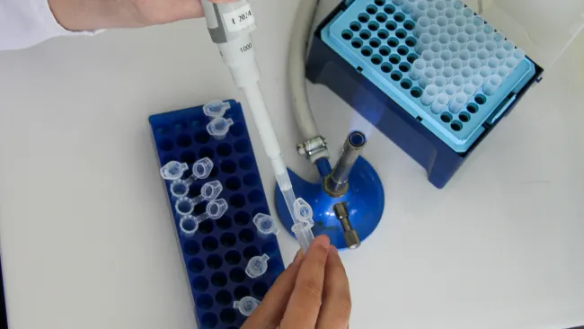
Prostate-Specific Antigen (PSA) Blood Test

PSA is a protein made by the prostate. Levels can rise with cancer, but they can also rise with infection, inflammation, BPH, and even recent activity. A single PSA result is just one piece of the overall picture. Your prior results, age, and prostate size all influence how the number is interpreted.

Digital Rectal Exam (DRE)

A brief physical exam that lets a urologist feel the back surface of the prostate for firmness, nodules, or asymmetry. It is not a substitute for PSA, and PSA is not a substitute for the exam; together they give a more complete picture.

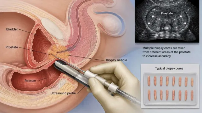
How Prostate Cancer Is Diagnosed

If a PSA result, a DRE finding, or a symptom raises concern, a urologist will usually take a stepped approach rather than jumping straight to a biopsy. The goal is to gather enough information to either rule out cancer or characterize it accurately enough to plan the right treatment.

Modern medical imaging equipment with digital monitors in a clean, white hospital examination room.

History and Exam

Your urologist will review your symptoms, your family history, your medications, and your overall health. The DRE is part of the same visit.

Scientist using a micropipette to transfer liquid into microcentrifuge tubes in a laboratory setting.

Repeat PSA and Additional Labs

A single elevated PSA is often re-checked, sometimes alongside other markers that help separate cancer-driven elevations from PSA elevations caused by BPH or inflammation.

Doctor preparing middle-aged man for MRI scan in modern medical imaging room with large scanner.

Imaging

Multiparametric prostate MRI has become an important step before many biopsies. It shows the prostate in detail and helps the team decide whether a biopsy is needed and, if so, where to focus.

Illustration of transrectal ultrasound guided prostate biopsy with labeled anatomy and biopsy procedure.

Prostate Biopsy

When a biopsy is the right next step, our team uses an MRI/ultrasound-guided transperineal approach. This technique targets suspicious areas identified on imaging and is performed through the skin between the scrotum and rectum, which can reduce certain infection risks compared to older approaches.

Microscopic view of clustered purple cells stained on a light pink tissue background under a microscope.

Pathology and Grading

Tissue from the biopsy is reviewed by a pathologist. The report names the type of cancer, assigns a Gleason score and Grade Group, and describes how much of each biopsy core was involved. That information, combined with PSA and imaging, builds the picture your treatment plan is based on.

Atlanta Prostate Center logo with blue geometric shapes and tagline The Future of Care Now

Call Atlanta Prostate Center Today

At Atlanta Prostate Center, prostate cancer care is all we do. If you or a loved one is facing a prostate cancer diagnosis, or you just want a second opinion, we are here to help you find the right path forward.

Treatment Options at a High Level

The right plan depends on the type of cancer, the grade, the stage, your age, and your overall health. Our role is to lay out the choices that fit your situation, explain the trade-offs, and let you decide with us.

Person administering insulin injection into abdominal fat for diabetes management
1

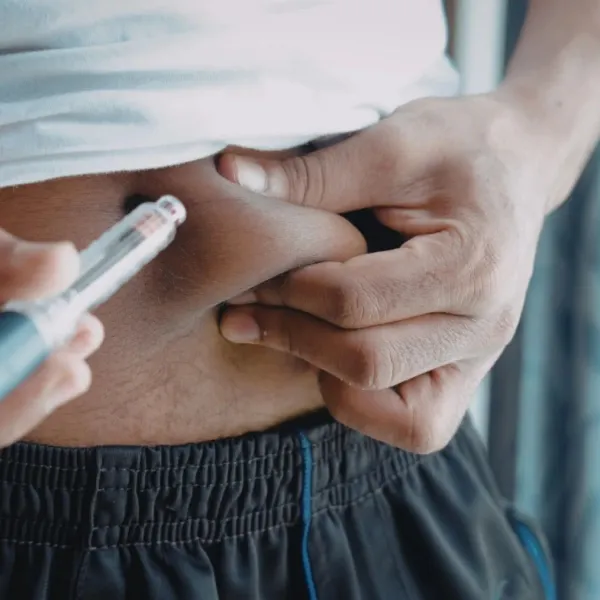
Androgen Deprivation Therapy

Prostate cancer cells rely on male hormones, primarily testosterone, to grow. ADT works by lowering hormone levels in the body or blocking them from reaching the cancer. It can be used on its own or paired with other treatments to improve outcomes.

Medical illustration of a male pelvic cross-section with a gloved hand pointing at the rectum anatomy.
2

Brachytherapy

Brachytherapy is a minimally invasive form of internal radiation therapy where tiny radioactive seeds are placed directly into the prostate. The radiation targets cancer cells from the inside while limiting exposure to surrounding healthy tissue.

Hands holding ultrasound probe and gel bottle in front of ultrasound machine monitor in medical setting.
3

HIFU (High-Intensity Focused Ultrasound)

HIFU uses sound waves to heat and destroy cancerous tissue within the prostate. Because the energy is targeted with extreme accuracy, surrounding healthy tissue is preserved, helping protect urinary and sexual function. HIFU is non-invasive, requires no incisions, and is performed as an outpatient procedure.

Advanced robotic surgical system operating in a modern hospital with monitoring screens and medical equipment.
4

Robotic Prostatectomy

Robotic prostatectomy is the surgical removal of the prostate using robotic-assisted technology, giving the surgeon enhanced precision, visualization, and control. The minimally invasive approach means smaller incisions, less blood loss, shorter hospital stays, and a faster recovery compared to traditional surgery.

Cross-sectional illustration of male pelvic anatomy showing bladder, prostate, seminal vesicles, and urethra.
5

Transperineal Prostate Biopsy

A transperineal biopsy uses MRI and ultrasound guidance to collect tissue samples through the perineum, the area between the scrotum and rectum, rather than through the rectal wall. This approach offers superior accuracy, better access to all regions of the prostate, and a lower risk of infection.

Close-up of a water droplet creating ripples on a clear blue water surface in soft lighting
6

Water Vapor Ablation

Water vapor ablation is a minimally invasive outpatient treatment that uses targeted bursts of steam to destroy unwanted prostate tissue. The thermal energy ablates the targeted area while preserving surrounding healthy tissue and protecting urinary and sexual function.

Gloved hand holding a blood sample tube labeled PSA Test with lab test results showing 12.6 H for PSA.
Two men smiling together indoors, one with arm around the other's shoulder, sitting on a couch by a window.
Man wearing a checkered shirt using a urinal in a restroom with wooden privacy dividers.
Elderly man with glasses sitting at a table, holding a fork with food, engaging in conversation indoors.

When to See a Urologist

Prostate cancer often develops without early symptoms, so knowing when to see a urologist matters; consider scheduling a visit if any of the following apply to you.

A higher-than-normal PSA result on a blood test can be an early indicator of prostate issues, and a urologist can help determine whether further evaluation or monitoring is needed.

If your father, brother, or other close relatives have been diagnosed with prostate cancer, your risk is significantly higher, and earlier screening with a urologist is strongly recommended.

Frequent urination, weak flow, difficulty starting or stopping, or blood in the urine can signal a prostate condition that warrants a professional evaluation.

Prostate cancer risk increases with age, so men 40 and older, especially those with added risk factors, should establish care with a urologist and discuss a screening schedule.