
A prostate MRI is one
of the most detailed and least invasive ways to get a close look at the
prostate. There is no surgery and no needle into the gland, just a series of
clear images.
What Is a Prostate MRI?
A prostate MRI is a
scan that uses a strong magnet and radio waves, not radiation, to create high-detail pictures of
your prostate and the tissue around it. Most practices use a version called
multiparametric MRI, often shortened to mpMRI. That simply means the scan
captures several different types of images, each one showing something a little
different about the gland. Put together, they give your urologist a clear view of the
prostate's size, shape, and any areas that look out of the ordinary. Some scans
add a contrast dye to highlight blood flow, which is why you may hear yours
described as an MRI with and without contrast.
Why Would a Doctor Order a
Prostate MRI?
An MRI for prostate
cancer is usually about answering one question: do we need to look closer, and
if so, where? Your doctor may order one for a few common reasons.
● A rising or elevated PSA on a blood test
● An area that felt firm
or irregular during a prostate exam
● Mapping the gland
before a biopsy so any suspicious spots can be targeted
● Keeping an eye on a
known low-risk cancer during active surveillance
●
Checking how the prostate looks after treatment
That last point about
targeting is a big deal. A good MRI can help your doctor decide whether a
biopsy is even needed, and when one is, it shows exactly where to aim.
How to Prepare for a
Prostate MRI
Prostate MRI
preparation is usually simple, and your imaging center will send you exact
instructions. Details vary from place to place, so always follow the steps your
center gives you. That said, here is what often comes up.
● You may be asked to use
a small enema beforehand so the rectum is empty and the images come out clearer
● Some centers ask you to
skip caffeine for a short window before the scan
● You will remove
anything metal, including jewelry, your watch, belts, and anything in your
pockets
● Tell the team about any
implants, a pacemaker, or metal fragments in your body, since these affect
whether an MRI is safe for you
●
Mention claustrophobia ahead of time, because a mild calming
medication can sometimes be arranged
What the Prostate MRI
Procedure Is Like
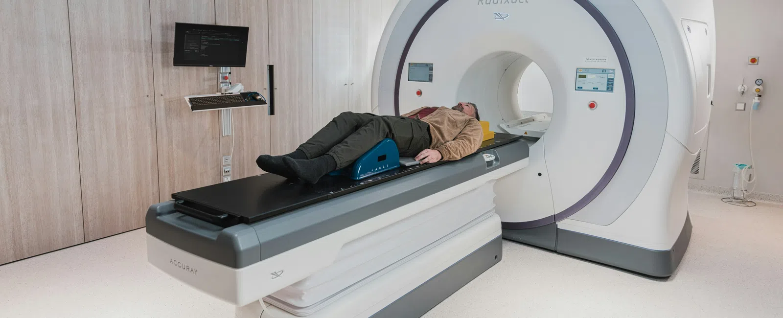
On the day, you change
into a gown and lie on your back on a padded table. The table slides into the
MRI machine, which looks like a wide tube that is open at both ends. The
scanner is loud, so you will get earplugs or headphones, and many centers will
even play music. Your main job is to stay still, since movement blurs the
pictures.
If contrast is part of
your scan, a small IV goes into your arm, and you might feel a brief cool
sensation when the dye is given. The prostate MRI procedure typically takes
somewhere around 30 to 45 minutes from start to finish. Older protocols
sometimes used a thin coil placed in the rectum for sharper images, though many
modern scanners no longer need this. You stay awake the whole time and can talk
to the technologist through a speaker whenever you need to.
Understanding Your Prostate
MRI Results 1 to 5
When a radiologist
reviews your scan, they often use a scoring system called PI-RADS, which stands
for Prostate Imaging Reporting and Data System. It rates how likely it is that
a suspicious area represents significant cancer, on a scale from 1 to 5. When
people search for prostate MRI results 1-5, this is almost always the scale
they are trying to understand.
● 1: very low likelihood
of significant cancer
● 2: low likelihood
● 3: uncertain, somewhere
in the middle
● 4: high likelihood
●
5: very high likelihood
Here is the important
context: a higher number does not mean you definitely have cancer, and a lower
number does not always rule it out. The score is one piece of a larger picture.
Your urologist reads it alongside your PSA, your exam, your family history, and your other risk
factors before deciding what comes next. If anything in your report looks
confusing, ask. It is your health, and you deserve a plain answer.
What Happens After the Scan?
If your MRI looks
reassuring and your PSA is steady, your doctor may simply recommend staying the
course with continued monitoring. For patients without a pre-existing diagnosis
of prostate cancer, it is important to note that a negative MRI (no PIRADS lesions
or low risk PIRADS lesions) does NOT rule out prostate cancer. Research shows
that approximately 20% of prostate cancers do not show up on MRI. Thus, in
patients with clinical features concerning for prostate cancer (elevated PSA),
a prostate biopsy is still recommended despite the MRI results.
If an area on the MRI
looks suspicious, your urologist will generally plan an MRI fusion biopsy, which blends your MRI
images with live ultrasound so the doctor can sample the exact spot of concern
rather than guessing. You can read how that compares with other approaches in
our breakdown of how a prostate biopsy is performed.
If a biopsy does find
cancer, your care team will walk you through how the Gleason score works and then the treatment options
available to you. Many of those paths are far less daunting than men expect, and
the right one depends entirely on your specific situation.
Talk With a Prostate
Specialist in Atlanta
A prostate MRI can feel like a heavy step, but really it is just a clear, careful look that helps you and your doctor make a smart decision together. If you have an elevated PSA or your primary care doctor has suggested imaging, the team at Atlanta Prostate Center can explain what your numbers mean and whether an MRI or a biopsy makes sense for you. Call us to schedule a consultation and get answers built around your situation, not a generic checklist.